Actin vs Myosin a Basic Interesting Difference Guide
Actin vs myosin, are two different protein types that exist in muscles and assist in muscle contraction in humans as well as in animals.
The difference in both these terms includes that actin manufactures thin contractable threads while myosin manufactures thick contractile threads within cells of muscles.
read: Humoral and cell-mediated immunity
These both are very important and basic components of muscle that help involuntary muscle movement management along with other proteins, for instance, tropomyosin, meromyosin and troponin.
Actin and myosin form threads which are managed longitudinal manner in the myofibrils. They are also employed for non-cellular and cellular movements.
let’s come to our main topic to explore difference between actin and myosin.
Difference between actin and myosin

Actin and myosin difference one by one:
| Actin | Myosin | |
| 1. | The filament size that actin forms is short and thin. | The filament size that myosin forms is long and thick. |
| 2. | It is located in A and I band. | It is located in A band of the sarcomere. |
| 3. | It possesses troponin and tropomyosin. | It possesses meromyosin. |
| 4. | Actin filaments are huge in numbers. | These are lesser in number and as 1 myosin filament is equal to 6 actin filaments. |
| 5. | The filaments of actin possess a smooth surface. | Filaments of actin possess rough surfaces. |
| 6. | It never forms cross-bridges. | It does form cross-bridges. |
| 7. | Actin filaments are one end free | Myosin filaments are both ends free. |
| 8. | At the contraction time, actin slides into H zone. | At the contraction time, it does not slide. |
| 9. | Actin possesses a smooth surface as compared to myosin. | It possesses a rough surface. |
| 10. | Filaments of actin are polymer of actin monomers, such filaments are the source for the creation of a core of thin muscle filaments. | These are motor molecules that utilize ATP to drag actin. |
Actin and myosin definition
Actin:
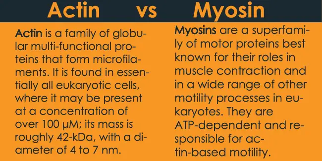
Actin is one of the fundamental ingredients of the cell cytoskeleton, particularly in eukaryotes. It is an exceptionally moderated protein having a sub-atomic load of 42kDa.
Actin is available in monomeric structure as G-actin or polymeric structure as F actin, where ‘G’ represent globular actin protein, while ‘F’ is for filamentous actin protein or polymeric stringy protein.
These have diverse cell capabilities like muscle withdrawal, cytokinesis, and cell relocation.
As actin filaments play a primary role in the formation of the dynamic cytoskeleton of the cell, hence it employs to provides the movement and shape also to the cell.
The cytoskeleton likewise conveys correspondences with the neighboring cells, additionally bolsters the inside encompassing within the cell.
Myosin:
Myosin is another filamentous protein type that employes in the calcium ion presence. Myosin is known to produce the energy that is a requirement during muscle withdrawal. In this way, the other name of the myosin protein is known as motor protein.
Skeletal muscles are known for the deliberate activity in the body, where actin and myosin are available as the rehashing units. The thick fiber of the myosin is encircled by the flimsy fiber of actions. Of course, the dainty fiber (thin fiber) of actin is encircled by the thick fiber of the myosin. Along these lines, this constant and rehashing bring about the arrangement of the heap of fibers in muscles.
Myosin has three sections: tail, head, and neck and is made out of various light chains and two substantial chains.
The globular head locale has the coupling site for the ATP and actin, and the neck part has alpha-helical district wheres tail has other restricted destinations. Head area changes over ATP to ADP, by the chemical ATPase.
When the nerve imparts sign muscle cell for the muscle withdrawal, the myosin and actin get in an active condition. From that point onward, the myosin begins working in discharging vitality (ATP), and further, the myosin along side actin fibers slide past one another.
Tropomyosin and troponin are the other 2 muscles protein types that brief with the actin and myosin for the muscle withdrawal. This working can be comprehended by the theory named as ‘sliding filament hypothesis’.
Actin and myosin additionally assume a basic job in nonmuscle cells. There are two activities performed successively by the muscles, which are compression and unwinding. The compression brings about the shortening of muscles and results in development while unwinding restores the muscle to its unique length.
Actin vs myosin similarities:
- Calcium particles/ions are required for the muscles constriction.
- The two kinds of proteins (actin and myosin) are required during muscle compression.
- Actin and Myosin are the protein fibers present in muscles.
Key actin myosin difference
- Actin and myosin are the proteins fibers found in muslces, and actin is known to frame the dainty groups in the myofibrils, while myosin is known to shape the thick groups in the myofibrils.
- Actin frames are tiny fiber of 2-2.6 um, and it is flimsy up to 0.005 µm, yet myosin shapes a long fiber of 4.5 µm, which has a thickness of 0.01 um, that implies actin are more slender than myosin.
- Actin possesses troponin and tropomyosin (proteins), and myosin contains meromyosin (protein), it(myosin) has an ATP restricting site, through which it discharges vitality for the muscle withdrawal.
- Actin is available in A & I groups, though myosin is available in A groups of the sarcomere.
- The outside of actin is smooth, and they are more in numbers when contrasted with the myosin, the proportion is one for every six actin particles. Myosin has an unpleasant surface.
- Actin slide in H zone at the time constriction, though myosin doesn’t slide at the hour of withdrawal.
Conclusion:
We can conclude that separated from the muscle constriction, actin and myosin assume a crucial job in cell science by taking its part in cell division, in elements of nonmuscle cells, and so forth. Myosin is thicker than actin and has darker striations. The working of muscle constriction can be comprehended by the sliding fiber hypothesis.
You may also enjoy reading:
Thank You very much Jane! keep coming back
Hey John! thank You 🙂